By Barry Keate
Barry Keate, has lived with tinnitus over 40 years and has published 150+ research articles on numerous aspects of tinnitus. He is an expert on the condition and a well-known advocate for those with tinnitus.
Author’s Note: Much of the information for this article was garnered from the American Academy of Otolaryngology-Head and Neck Surgery Foundation (AAO-HNSF), especially the book “Primary Care Otolaryngology” by Gregory Staffel, MD, who donated the book to the AAO-HNSF. Other material came from the Mayo Clinic and the American Speech-Language-Hearing Association. We are indebted to these organizations for their contributions.
Tinnitus is most frequently the result of hearing loss and most people who experience hearing loss will have tinnitus as one of the symptoms. While exact numbers are difficult to determine, the American Tinnitus Association estimates that 70% of tinnitus is due to hearing loss. This overview will discuss the various types of hearing loss, the causes and available treatments, when applicable.
Hearing Loss
The ear consists of three major areas: the outer ear, middle ear and inner ear. In normal hearing, sound vibrations are funneled by the outer ear into the ear canal where they cause vibrations in the eardrum. These vibrations transfer to the three small bones of the middle ear, the malleus (hammer), incus (anvil), and stapes (stirrup), which amplify the vibrations as they travel to the inner ear. There, the vibrations pass through fluid in the cochlea, a snail-shaped structure in the inner ear. Attached to nerve cells in the cochlea are thousands of tiny hairs that help translate sound vibrations into electrical signals that are transmitted to the brain. The vibrations of different sounds affect these tiny hairs in different ways causing the nerve cells to send different signals to the brain so it can distinguish one sound from another.
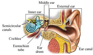
There are two basic types of hearing loss: conductive hearing loss, and sensorineural hearing loss. Sometimes there are elements of both and it is termed mixed hearing loss. Conductive hearing loss occurs when sound is not conducted efficiently through the outer ear canal to the eardrum and the small bones of the middle ear. The most prevalent causes of conductive hearing loss are: fluid in the middle ear from colds, allergies, eustachian tube dysfunction, ear infection; otosclerosis; perforated eardrum; and impacted earwax.
Sensorineural hearing loss occurs when there is damage to the inner ear, or cochlea, or to the nerve pathways from the inner ear to the brain. This accounts for the majority of hearing loss. Sensorineural hearing loss is considered by the medical establishment to be permanent because there is no medically recognized treatment or surgery that will cure the condition.
The most prevalent causes of sensorineural hearing loss are: disease; drugs that are toxic to the auditory system (ototoxic); noise exposure; viruses; head trauma; aging; and tumors.
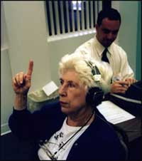 During the research for this article I was intrigued to discover how the various tests for hearing loss, audiograms and tympanograms, can narrow down the type of hearing loss and provide very precise information on exactly what problems may have developed and how well the ears are functioning. Pure tone audiometry is used to assess the patient’s hearing levels. During the audiogram, independent hearing thresholds are determined for both air conduction and bone conduction. Air conduction is when the sound travels through the air into the ear and the cochlea. This measures the ability of the ear to conduct sound. Bone conduction bypasses the middle and outer ear by sending sound waves through the mastoid bone directly to the cochlea. This tests for sensorineural hearing loss.
Tympanograms test for mobility of the ear drum which can determine whether there is high or low pressure in the middle ear, caused by fluid build-up or negative pressure due to poor eustachian tube function.
Speech discrimination testing is also conducted by presenting phonetically similar sounds into the audiogram. This test of clarity also reveals the function of the auditory, or 8th cranial, nerve. Amplifying garbled speech with a hearing aid has very little benefit for someone with poor speech discrimination.
Hearing threshold levels are determined between 250 and 8000 hertz (Hz) and measured in decibels (dB). Human speech ranges from 300 to 4,000 Hz. The 0 dB level is normalized to the minimum hearing level of young healthy adults and does not mean there is an absence of sound. The sound level is increased at each frequency until it is heard by the patient. The higher the threshold level, the poorer the patient’s hearing. Thresholds higher than 25 dB are considered abnormal.
Figure 6.1 demonstrates the precision and versatility of an audiogram. Note that the left ear has normal bone conduction but decreased air conduction. This indicates a fluid build-up in the left ear that is impairing hearing.
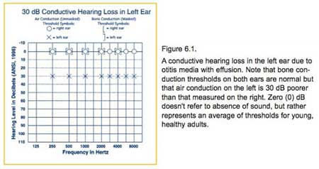
Figure 6.2 shows a typical audiogram for someone with age-related hearing loss (presbycusis).
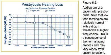
Figure 6.3 shows a typical audiogram of someone with noise-induced hearing loss. Note the typical notch of decreased hearing at 4,000 Hz.
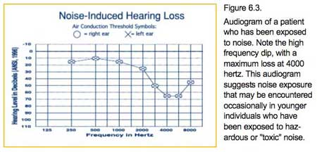 Author’s Note: This is the type of hearing loss that many tinnitus sufferers and I display. My first audiogram 35 years ago was nearly identical to this.
Conductive Hearing Loss
There are many contributing factors to conductive hearing loss, many of which impact directly on tinnitus. Most of these factors are treatable and we will describe each in turn.
Otitis media refers to inflammation of the middle ear and may be thought of in terms of eustachian tube dysfunction. This can occur due to a cold, upper respiratory infection or allergy. The eustachian tube becomes obstructed resulting in negative pressure in the middle ear. This, in turn, forces fluids to pass through the membranes and fill the middle ear. When infection occurs in these fluids, it is called acute otitis media. The infection is usually the result of Streptococcus pneumonia and is treated with antibiotics. When the middle ear is filled with fluid that is not infected, it is termed otitis media with effusion. Treatment for otitis media without infection can be as simple as a prescription nasal spray, such as Flonase, and taken with an antihistamine.
Many children are susceptible to acute otitis media and may have several episodes. These children will often benefit from a pressure equalization tube inserted through the eardrum to vent the middle ear and prevent negative pressure. It is important to note that the tube is not intended to drain the fluid but is for pressure equalization. Children often grow out of the eustachian tube dysfunction by the time the tubes fall out on their own, in about 1-2 years.
All people with middle ear fluid or infection have some degree of hearing loss. The average hearing loss in otitis media with effusion is 24 dB, equivalent to wearing earplugs, however thicker fluid can cause hearing loss up to 45 dB.
Otosclerosis is a hereditary condition of abnormal bone growth in the tiny bones of the middle ear. This leads to a fixation of the stapes bone. The term is derived from the Greek “sclero” (hard) and “oto” (ear).
It is estimated that 10% of the adult Caucasian population is affected by some degree of otosclerosis. The primary symptom is slowly progressing hearing loss that can begin anytime between the ages of 15 and 45 but it usually starts in the early 20’s. Tinnitus is a frequent result of otosclerosis.
There is a surgical therapy for otosclerosis called a stapedectomy. This involves removing the immobilized stapes bone and replacing it with a prosthetic device. The device allows the bones of the middle ear to resume movement, which stimulates the fluid in the inner ear and improves or restores hearing. You can read more about stapedectomy in our Tinnitus Library.
Perforated eardrum can occur if the ear is struck squarely creating a pressure trauma. Other common causes are explosions, skull fractures, objects piercing the eardrum and untreated acute otitis media. On rare occasions a small hole may remain after a previously placed pressure equalization tube is removed or falls out.
Most eardrum perforations heal by themselves within weeks, although some may take up to several months. During healing the ear must be protected from water and trauma. If water leaks through the perforation, infection can occur.
Usually, the larger the perforation is, the greater the loss of hearing. The location of the perforation also affects the degree of hearing loss. If the perforation is due to a sudden traumatic or explosive event, the hearing loss and resultant tinnitus can be severe. In this case the hearing usually partially returns and tinnitus diminishes in a few days.
If the perforation fails to heal on its own an ENT physician may try to patch the eardrum in the clinic. The doctor will apply a chemical to the edges of the tear to promote growth and then place a thin paper patch over it. Usually an improvement in hearing is noticed immediately. Several applications of the patch may be necessary to completely heal the rupture.
If the paper patch method fails, a surgery called a tympanoplasty is usually performed. This involves placing living tissue over the perforation and letting it grow into the rest of the tissue. Surgery is usually very successful in permanently closing the perforation and improving hearing.
Impacted earwax is a common cause of hearing loss and tinnitus. The ear canal is shaped like an hourglass, narrowing part way down. The skin of the outer part of the canal has glands that produce earwax. Wax is not formed in the deep part of the ear canal. The wax is there to trap dust and dirt and keep them from reaching the delicate eardrum. The wax often accumulates, dries out and falls out of the ear, carrying dirt and dust with it. This is healthy in normal amounts and also coats the skin of the ear canal and acts as a water repellant. The absence of earwax results in dry, itchy ears.
The ear canal may be blocked by wax when attempts to clean the ear push the wax deeper into the canal and cause a blockage. When a person has earwax blocked against the eardrum, it is most often because he or she has been probing the ear with such things as cotton-tipped applicators, bobby pins or twisted napkin corners.
Most cases of earwax build-up respond to home treatments used to soften wax if there is no hole in the eardrum. Applying commercial earwax removal drops such as Mack’s Wax Away, Murine, or Physicians’ Choice will soften the wax; or applying a few drops of mineral oil, baby oil, or glycerin. The ear canal can then be flushed with hydrogen peroxide or rubbing alcohol.
In the event the home treatments are not satisfactory, or if the wax has accumulated to the extent that it blocks the canal and reduces hearing, a physician may describe eardrops designed to soften wax, or he may wash or vacuum it out. Occasionally an ENT specialist may need to remove the wax using microscopic visualization.
Author’s note: I have heard from more than a few people who had horrible experiences having earwax vacuumed out. In the hands of an inexperienced doctor, this can lead to a worsening condition and tinnitus can be dramatically increased. I do not recommend vacuuming earwax.
Sensorineural Hearing Loss
There are many contributing factors to sensorineural hearing loss. Most of these are considered untreatable by the traditional medical establishment because there are no medically recognized therapies or surgeries that will cure the condition.
Despite that opinion, there are many treatment options that, while not a cure, will result in a lessening of the tinnitus associated with hearing loss. These treatments range from diet and exercise, to supplements, sound therapy and some prescription medications. Most of these treatment options can be seen in our Tinnitus Library .
Disease Conditions
Disease conditions such as Meniere’s disease can lead to sensorineural hearing loss and tinnitus. Little is known about the underlying cause of Meniere’s disease however there are treatments for it. It involves a fluid build-up in the vestibular system that will eventually damage the hair cells of the cochlea leading to permanent hearing loss and tinnitus. A complete discussion of Meniere’s disease can be seen in our Tinnitus Library.
Thyroid disease
Thyroid disease usually causes hearing impairment and tinnitus. The condition results in a decrease in thyroid hormones that can also cause Fibromyalgia and Chronic Fatigue Syndrome. This condition is commonly treated with prescription thyroid hormones. A full discussion on thyroidism can be seen in our Tinnitus Library.
Ototoxic Drugs
There are over 200 prescription and over-the-counter medications that can cause or worsen hearing loss. We have heard from countless people who complained of hearing loss and tinnitus after taking a new medication. Anyone who already has hearing loss should exercise caution when taking new prescription medications. Make certain your physician knows about your hearing condition and concerns. An article on ototoxic medications can be seen in our Tinnitus Library .
Noise Exposure
This is a very common cause of hearing loss and tinnitus and is the cause of my hearing loss, as mentioned in the previous article. Noise exposure permanently damages the hair cells in the cochlea. It is common in certain industries and is closely regulated by the Occupational Health and Safety Administration (OSHA).
The following graph compares the loudness of common sounds.
|
Sound levels of common noises
|
|
|
Decibels
|
Noise source
|
|
30
|
Whisper
|
|
60
|
Normal conversation
|
|
90
|
Heavy traffic, garbage disposal
|
|
Risk Range
|
|
|
85 to 90
|
Motorcycle, snowmobile, lawn mower
|
|
90
|
Belt sander, tractor
|
|
95 to 105
|
Hand drill, bulldozer, impact wrench
|
|
110
|
Chain saw, jack hammer
|
|
Injury range
|
|
|
120
|
Ambulance siren
|
|
140
(pain threshold) |
Jet engine at takeoff
|
|
165
|
Shotgun blast
|
|
180
|
Rocket launch
|
Below are the maximum noise levels on the job to which you should be exposed without hearing protection and for how long. Most experts agree that continual unprotected exposure to more than 85 decibels is dangerous and leads to hearing loss.
|
Maximum job-noise exposure allowed by law
|
|
|
Decibels
|
Duration, daily
|
|
90
|
8 hours
|
|
92
|
6 hours
|
|
95
|
4 hours
|
|
97
|
3 hours
|
|
100
|
2 hours
|
|
102
|
1.5 hours
|
|
105
|
1 hours
|
|
110
|
30 minutes
|
|
115
|
15 minutes
|
Presbycusis
Commonly referred to as age-related hearing loss, Presbycusis is by far the most frequent cause of hearing loss in the elderly. As we age, the outer hair cells in the cochlea gradually deteriorate causing bi-lateral hearing loss, primarily in the higher frequencies. Patients with presbycusis may also have difficulty with speech discrimination and complain of tinnitus.
One of the most widely investigated potential causes of presbycusis concerns reduced blood flow in the cochlea associated with age that contributes to the formation of oxygenated free radicals. These molecules damage the mitochondrial DNA that leads to problems with neural functioning in the inner ear.
 Presbycusis can be prevented but once it occurs it joins the stable of other causes of sensorineural hearing loss and becomes permanent. Michael Seidman, MD has done pioneering work in this area and has obtained a US patent for a product that prevents mitochondrial damage to the inner ear. The product is called the Anti-Age/Energy Formula.
Dr. Seidman has also written a book called “Save Your Hearing Now” that details the progressive damage done to the inner ear by free radicals and outlines a complete plan for preventing damage and prolonging acute hearing ability.
Tumors
The primary tumor associated with hearing loss is an acoustic neuroma. This is a very rare, slow-growing, non-malignant tumor that occurs on the 8th cranial nerve controlling hearing and balance. In many cases these are left alone, especially if the patient is elderly and the tumor small. In other cases they must be surgically removed. If left to grow too large they will eventually impact on the brain and can be life threatening. Read a complete discussion on acoustic neuroma in our Tinnitus LIbrary.
Head Trauma
Numerous reports in the literature indicate that head trauma, which includes concussion and whiplash, causes hearing loss and tinnitus. Damage can occur to the bones of the middle ear or the 8th cranial nerve. The hearing loss may be temporary or permanent, depending on the degree of damage. The hearing loss mimics the hearing loss due to noise exposure, with a typical downward notch at 4 KHz.
Arches Tinnitus Formulas were developed to help people suffering from tinnitus due to sensorineural hearing loss, regardless of the cause. While not a cure, the formulas have helped thousands of people reduce the sound level and continue with an enjoyable life. Read a highly informative article on the Science Behind the Product .
